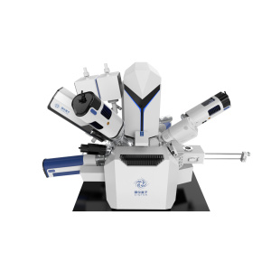
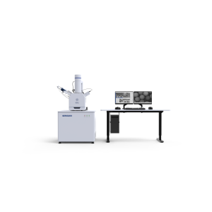
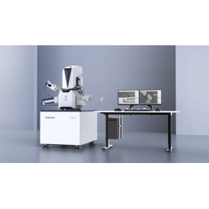
A scanning electron microscope (SEM) produces images of a sample by scanning the surface with a focused beam of electrons. The signals that derive from electron-sample interactions reveal information about the sample including morphology, chemical composition, and crystalline structure and orientation of materials making up the sample.
SEMs can achieve resolutions down to 1 nanometer.
It can be combined with a wide variety of detectors providing different information on the samples:
1) Morphology, texture and discrimination of phases with SED and BSD .
2) Elemental mapsor spot chemical analyses using EDS.
3) Compositional maps based on differences in trace element “activitors” (typically transition metal and Rare Earth elements) using CL.
4) Crystallographic information using EBSD